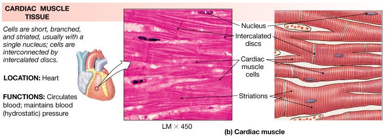
Label The Features Of Cardiac Muscle Tissue
Why are cardiac muscles so called? explain their structure with the Muscle cardiac tissue microscopic anatomy photomicrograph fibers intercalated accompanying physiology striations reviewing nucleus 775x Cardiac histology labeled microscope cells
Cardiac muscle (labeled) | Karen Reeds | Flickr
Cardiac muscle 40x intercalated discs histology Cardiac muscle tissue Cardiac muscle (labeled)
Cardiac muscle intercalated gap discs tissue junctions sarcolemma heart structure junction desmosomes figure desmosome fiber anatomy labeled they part shows
Cardiac muscle 40xCardiac muscle cells muscles tissue diagram cell muscular smooth model system animal tissues drawing gif wordpress filament skeletal function anatomy Cardiac muscle tissue heart cell skeletal striated forensics kenya resource onlineHistology-world! histology fact sheet-heart histology.
Histology of cardiac muscleAnatomy and physiology archive Cardiac muscle section stain tissue longitudinal human plate hematoxylin acid zenker fluid anatomyatlasesCardiac muscle electrical activity cells intercalated structure labeled discs anatomy cross physiology parts myofibrils.

Histology muscle cardiac slides slide labels heart myocardium parts world microscope endocardium pathology photomicrographs another website epicardium visit album
Physiology glossary: cardiac muscle cellCardiac labeled farm8 Kenya forensics online resource: cardiac muscle tissueCardiac cell muscle anatomy draw cellular.
Muscular tissueSolved: label the structures found in cardiac muscle tissue. Muscle cardiac label structures tissue found show transcribed textPlate 5.76: cardiac muscle.

Cardiac labelled mammalian
Cardiac muscle and electrical activity .
.